Three-Compartment Breast Lesion Detection (3CB)
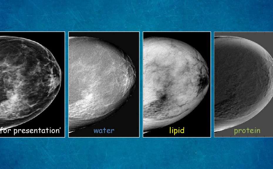
The Shepherd Research Lab has developed a novel breast-imaging technique that analyzes lesion composition using three compartmental measurements: protein, lipid, and water (3CB). The study investigates whether lipid-protein-water signatures of mammographically suspicious breast lesions can improve cancer diagnosis and reduce unnecessary biopsies.
The first five years of research (March 2013 – June 2017) under NCI R01CA166945 established the imaging method and produced the publications that supported a competing renewal. The renewal — NCI R01CA257652 (08/09/2021 – 07/31/2026) — extends 3CB by combining the compositional biomarkers with quantitative image analysis (QIA) and radiomics, collectively termed q3CB.
Long-term goals
- Determine whether biological diagnostic measures can improve CADe (computer-aided detection) algorithms
- Quantify lipid-protein-water signatures to predict malignant findings
- Combine 3CB biomarkers with existing QIA/radiomics methods to improve sensitivity and specificity
- Reduce unnecessary biopsies
Aim 1 — Sensitivity & specificity of 3CB signatures
Recruitment adjusted from 600 to 498 FFDM patients due to a 17% budget reduction. As of the last update, 425 women enrolled (215 UCSF / 210 Moffitt) with biopsy-confirmed subtypes: 61 invasive ductal carcinoma (IDC), 40 ductal carcinoma in situ (DCIS), 66 fibroadenomas, 324 benign findings.
A calibration phantom with 51 combinations of water, lipid (wax), and protein (Delrin) was created for standardizing 3CB across FFDM and DBT systems.
Early analysis of 45 lesions demonstrated 3CB features distinguishing between lesion types:
- IDC from DCIS by lipid skewness — AUC = 0.71
- Fibroadenomas by water texture relative to background — AUC = 0.75
- Benign lesions by peripheral water content — AUC = 0.71
Combined 3CB signature achieved AUC = 0.72 in cross-validation for invasive-cancer detection.
Aim 2 — Comparison with CAD/QIA methods
Merged QIA/radiomics signatures yielded AUC = 0.81 in distinguishing lesions requiring biopsy from those not requiring it. Key QIA features identified:
- Invasive cancer: spiculation
- DCIS: circularity
- Fibroadenoma: radial gradient index
- Other benign findings: texture heterogeneity
Deep-learning approaches for microcalcification classification showed promising results. In a dataset of 99 biopsy-proven lesions, the deep-learning method “could have avoided 21 biopsies of the 80 benign lesions…versus only 8 avoidable biopsies based on radiologists (p < .001).”
Aim 3 — Combined 3CB and QIA performance
The combination of 3CB and QIA (q3CB) significantly improved classification:
| Lesion type | 3CB alone | QIA alone | Combined |
|---|---|---|---|
| Overall | 0.71 | 0.81 | 0.86 |
| Masses | — | 0.83 | 0.89 |
| Microcalcifications | — | 0.84 | 0.91 |
| Asymmetry / arch. distortion | — | 0.61 | 0.87 (p = 0.006) |
3CB compositional information and QIA features provide complementary diagnostic information with little correlation between them.
Future directions
Extension to 3D tomosynthesis imaging and reader studies to validate whether 3CB knowledge influences radiologist decision-making and reduces unnecessary biopsies.
Funding
NCI R01CA257652 — Lesion Composition and Quantitative Imaging Analysis on Breast Cancer Diagnosis
Competing renewal · 08/09/2021 – 07/31/2026 · MPI: John A. Shepherd (contact, UH Cancer Center) and Maryellen L. Giger (University of Chicago). NIH RePORTER ↗
Project summary. Women with dense breasts have not been shown to benefit from increased cancer detection by volumetric digital breast tomosynthesis (DBT) but may benefit from lower recall rates. DBT screening biopsy rates are similar to 2D digital mammography — higher for first screening exams, lower thereafter, after adjusting for age and breast density. In the U.S., 71% of biopsies do not result in a breast cancer diagnosis among women ages 40–79 who undergo breast cancer screening. To address this high rate of unnecessary biopsies, the project uses FDA-approved breast imaging protocols to acquire multispectral images that measure the lipid/water/protein (L/W/P) composition of suspicious breast lesions. Malignant breast tissue has unique L/W/P composition fractions compared to normal or benign breast tissue.
The proposal aims to increase biopsy yield (BI-RADS-PPV3) by combining L/W/P biological biomarkers with quantitative morphological and textural image analysis — together called q3CB. The benefits of adding q3CB to the current DBT screening/diagnostic imaging paradigm, which may already include computer-aided detection, are not yet known. The central hypothesis is that biological L/W/P fractions in breast tissue, in combination with morphological and textural tissue characteristics, will yield significantly higher breast cancer specificity than conventional interpretation of DBT alone. The long-term goal is to reduce unnecessary biopsies and increase biopsy yield.
Specific aims:
- Develop q3CB lesion signatures for distinguishing breast cancer lesions from benign lesions, using 600 prospectively-acquired DBT exams of women recommended to undergo biopsy.
- Conduct a clinical reader study to compare radiologists’ performance on standard-of-care FFDM or DBT without and with the inclusion of q3CB signatures.
- Investigate the utility of q3CB lesion signatures in a screening paradigm to improve sensitivity and specificity on CADe-identified suspicious lesions — both for assessing malignancy and for association with cancer subtypes.
- Exploratory: explore the added sensitivity and specificity of dual-energy DBT in phantom studies that vary lesion size, composition, and breast density.
The innovation of the study is the full characterization of lipid/water/protein lesion composition with DBT and how it complements existing computer-aided diagnostic programs paired with clinical radiologists, providing evidence ready for clinical translation.
NCI R01CA166945 — Lesion Composition and Quantitative Imaging Analysis on Breast Cancer Diagnosis
Original award · 03/06/2013 – 02/28/2019 · MPI: John A. Shepherd (contact) and Maryellen L. Giger. Began at UC San Francisco; transferred to the UH Cancer Center in 2017 with a one-year extension.
General Electric — Investigator-Sponsored Research Contract
08/01/2018 – 07/31/2020.
Key publications
-
Leong LT, Malkov S, Drukker K, et al. Dual-energy three-compartment breast imaging for compositional biomarkers to improve detection of malignant lesions. Communications Medicine. 2021 Aug 31;1(1):29. DOI: 10.1038/s43856-021-00024-0
-
Hinton B, Ma L, Mahmoudzadeh AP, et al. Derived mammographic masking measures based on simulated lesions predict the risk of interval cancer after controlling for known risk factors. Med Phys. 2019 Mar;46(3):1309–16. DOI: 10.1002/mp.13410
-
Drukker K, Giger ML, Joe BN, et al. Combined benefit of quantitative three-compartment breast image analysis and mammography radiomics in the classification of breast masses in a clinical data set. Radiology. 2019;290(3):621–8. DOI: 10.1148/radiol.2018180608
-
Gierach GL, Patel DA, Falk RT, et al. Relationship of serum estrogens and metabolites with area and volume mammographic densities. Hormones and Cancer. 2015;6(2–3):107–19. DOI: 10.1007/s12672-015-0216-3
-
Malkov S, Kerlikowske K, Shepherd J. Automated volumetric breast density derived by shape and appearance modeling. Proc SPIE Int Soc Opt Eng. 2014 Mar 22;9034:90342t. DOI: 10.1117/12.2043990