Deep Learning and Total-Body DXA Scans (TBDXA.I.)
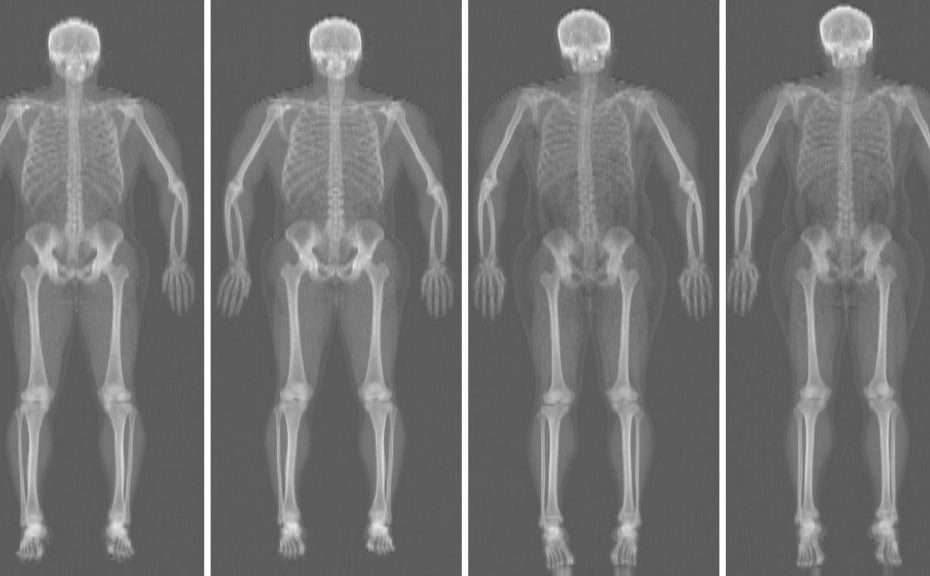
This research applies deep-learning methods to total-body DXA (dual-energy X-ray absorptiometry) scans to extract predictive information about body composition and health outcomes. The project uses self-supervised learning on whole-body images from the Health, Aging and Body Composition (Health ABC) Study, which includes over 3,000 participants with follow-up scans at years 3, 6, and 10.
The approach generates algorithms to predict clinical outcomes — cardiovascular disease, mortality, cancer, hip fracture, physical disability, diabetes-related measures — extracting features from unlabeled DXA images without requiring labeled outcome data during the initial learning phase.
Specific aims
- Predict clinical endpoints — generate predictive algorithms for CVD, CVD death, overall mortality, cancer, cancer death, hip fracture, physical disability, incident insulin-resistant diabetes, and metabolic markers by applying deep learning to baseline and follow-up Total-Body DXA images.
- Predict physical performance and inflammation — algorithms for gait speed, corridor walk speed, and IL-6 concentration.
- Explain the model — explore image features accounting for algorithm accuracy using saliency mapping techniques.
Research team
Principal Investigators
- Steve Cummings, MD, FAPC — San Francisco Coordinating Center
- John Shepherd, PhD — UH Cancer Center
Co-Investigator
- Peter Sadowski, PhD — UH Mānoa, Information and Computer Sciences
Additional team members
- Warren Browner, MD, MPH — CEO, California Pacific Medical Center
- Eleanor Simonsick, PhD — Epidemiologist, National Institute on Aging (NIH)
- Yannik Glaser — Graduate Student, UH Mānoa ICS
- Lily Liu — Statistician, California Pacific Medical Center
Funding
Sutter Health / California Medical Center Research Institute — Grant 2805096-0100 · 11/01/2019 – 10/31/2020.
Key publication
Glaser Y, Shepherd J, Leong L, Wolfgruber T, Lui L-Y, Sadowski P, et al. Deep learning predicts all-cause mortality from longitudinal total-body DXA imaging. Communications Medicine 2022 Aug 16;2. DOI: 10.1038/s43856-022-00166-9
The strongest model achieved an area under the ROC curve of 0.79 on held-out test data from over 15,000 scans. The study employed explainable-AI techniques to interpret predictions and evaluate input contributions.
Presentation
Glaser Y, Sadowski P, Wolfgruber T, Lui L-Y, Cummings S, Shepherd J. Hip fracture risk modelling using DXA and artificial intelligence. Poster, American Society for Bone Mineral and Research Annual Meeting; 2020 Sep 11; Virtual.